· High-quality Fluent Imaging (HQFI): Unique image algorithms and powerful image post-processing functions meet all digital clinical diagnosis requirements.
· Noise Suppression and Enhancement Based on Tissue Density and Morphology (DPE): The integrated tissue and morphology-based image enhancement technology captures high-quality images with clearer details and finer organization, ensuring easier visualization of subtle lesion tissues.
· Diagnostic Vision Optimization (DOO): This increases the visual comfort when viewing the image and reduces visual fatigue, while ensuring the quality of image diagnosis.
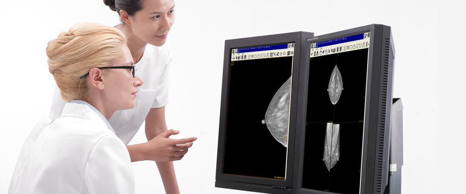
The independently developed integrated workstation can quickly process scanning operations and post-adjustment of images. The preset work interfaces may be accessed with one key operation. The system supports multi-user concurrent operation. The quick queries are customizable, and reports can be edited using a WYSIWYG mode.
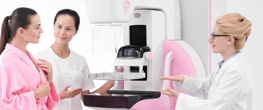
The UI is easy to use and intuitive, with more user-centric operation design.
The system seamlessly interfaces with the international standard DICOM protocol interface, and supports interconnectivity with RIS, PACS, HIS data.