The hardware and software not only work well together, but also complement each other. With free lifetime software updates, your device will never be out of date.
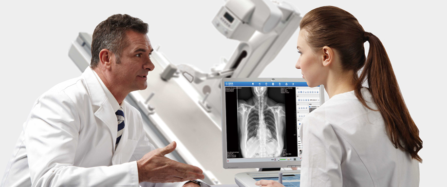
With nearly 20 years of medical imaging software development experience, our independently developed integrated workstation can quickly process scanning operations and post-adjustment of images. The preset work interfaces can be accessed with the press of a button. The system supports concurrent multi-user operation. The quick queries are customizable, reports can be edited using a WYSIWYG mode.
The brand-new upgraded UI is easy and intuitive. The graphical menu further streamlines the operation. Perspective and film modes may be switched with the press of a button, greatly optimizing the workflow.
The system seamlessly interfaces with the international standard DICOM protocol interface, and supports interconnectivity with RIS, PACS, HIS data.


Urinary system: IVP direct digital overall imaging can generate panoramic images of the urinary system
Real-time digital imaging of abdominal plain film
Gastrointestinal system: Long axis of the esophagus, panoramic view of the stomach, partially enlarged digital image of small sulcus and gastric area, panoramic view of the lower digestive tract
Respiratory system: Both, chest DR and direct digital chest radiograph under fluoroscopy
Orthopedics: High-definition bone imaging, joint imaging, long-axis imaging (judgment after overall healing)
Nervous system: Micro imaging of intracranial ear, temporal bone and paranasal sinuses
Peripheral blood vessel: Venous venography of limbs
Special angiography: Spinal cavity, salivary canal, uterine fallopian tube, etc.
Immune endocrine system: Direct observation of gout, rheumatoid bone and joint lesions, with sharper images
Emergency diagnosis: Rapid imaging, rapid positioning under fluoroscopy
Photography: Supports digital photography of anatomical parts of the whole body